Key takeaways
- Mitochondrial-derived peptides (MDPs) are short peptides encoded by small open reading frames hidden inside the mitochondrial 12S and 16S ribosomal RNA genes.
- Humanin, discovered in 2001, binds the pro-apoptotic protein Bax and the growth-factor binding protein IGFBP-3; its circulating levels fall with age in humans and mice.
- The six small humanin-like peptides (SHLPs) and MOTS-c were characterized in 2015 and 2016 by Pinchas Cohen’s group, and MOTS-c has since been shown to move into the nucleus under stress.
- Nearly all functional evidence comes from cell culture and rodents; no MDP has been tested as an intervention in a controlled human trial.
For most of the twentieth century the mitochondrial genome was thought to be fully accounted for. Its 16,569 base pairs encode 13 protein subunits of the respiratory chain, 22 transfer RNAs and two ribosomal RNAs, and the map appeared complete. Then, in 2001, a Japanese group hunting for factors that could protect neurons from Alzheimer-related damage pulled out a sequence that mapped not to the nuclear genome but to the mitochondrial 16S rRNA gene. The peptide it encoded was named humanin. Over the following fifteen years, researchers found a second family of six peptides in the same gene and a further peptide, MOTS-c, in the 12S rRNA gene. Together these are the mitochondrial-derived peptides, and they have changed how biologists think about what mitochondria do beyond producing ATP. This note surveys the family, the evidence for each member, and the gaps.
What makes a peptide “mitochondrial-derived”
The defining feature is genomic origin. MDPs are translated from short open reading frames (sORFs) embedded in mitochondrial DNA, in regions that also serve as templates for ribosomal RNA. Because the mitochondrial and cytoplasmic translation systems use different genetic codes, a given sORF can yield slightly different peptides depending on where it is translated; humanin is 24 residues if made in the cytoplasm and 21 if made inside the mitochondrion.3 Where MDP translation actually occurs remains under study, with evidence that transcripts are exported and translated on cytoplasmic ribosomes.
The concept matters because it implies a retrograde signaling channel. Mitochondria are known to influence the nucleus through metabolites, reactive oxygen species and calcium. MDPs add a class of peptide messengers that the organelle’s own genome encodes, whose levels can change with cellular stress and with age.9
Humanin
Hashimoto and colleagues identified humanin in 2001 by screening a cDNA library from the surviving occipital lobe of a patient with Alzheimer disease for sequences that protected cultured neurons against death induced by mutant amyloid precursor protein and presenilin genes. The protective clone encoded a 24-amino-acid peptide whose sequence lay within the mitochondrial 16S rRNA gene. Synthetic humanin protected neurons from a range of Alzheimer-related insults in vitro, and a serine-to-glycine substitution at position 14 (the analog known as HNG) was roughly a thousandfold more potent.1
Two mechanisms were described within two years. Guo and colleagues showed that humanin binds the pro-apoptotic protein Bax in the cytoplasm and prevents it from translocating to mitochondria, where it would otherwise trigger cytochrome c release and cell death.2 Ikonen and colleagues found that humanin also binds insulin-like growth factor binding protein 3 (IGFBP-3), a protein that modulates IGF-1 signaling and can itself induce apoptosis.10 Later work identified cell-surface receptors, including a trimeric complex involving the cytokine receptor gp130, though the full receptor biology is still being worked out.3
Humanin’s connection to aging came from population and animal studies. Yen and colleagues reported in 2020 that circulating humanin declines with age in humans and in mice, while the naked mole-rat, a species with negligible senescence, maintains stable levels. Offspring of centenarians had markedly higher circulating humanin than age-matched controls. In middle-aged mice, the HNG analog improved metabolic healthspan measures and lowered inflammatory markers.7 These are associations and rodent interventions; humanin has not been given to people in a controlled trial.
The small humanin-like peptides
In 2016 Cobb and colleagues searched the 16S rRNA gene systematically for additional sORFs and characterized six, naming them small humanin-like peptides 1 through 6 (SHLP1–6). The peptides ranged from 20 to 38 amino acids and were detected in mouse and human tissues and plasma. SHLP2 and SHLP3 were the most active: in cultured cells they reduced apoptosis and reactive oxygen species generation, promoted pre-adipocyte differentiation and improved mitochondrial oxygen consumption. In rats, SHLP2 infused into the brain increased peripheral glucose uptake and suppressed hepatic glucose output, an insulin-sensitizing profile acting at both central and peripheral levels. Circulating SHLP2 fell with age, as humanin does.5 SHLP6, by contrast, promoted apoptosis in the cell lines tested, a reminder that the family is not uniformly protective.
The SHLPs are the least studied members of the family. Their receptors are unknown, their tissue distribution is partly characterized, and the 2016 paper remains the primary source for most claims about them.
MOTS-c
MOTS-c is the only MDP encoded in the 12S rRNA gene and the only one that has reached even early clinical development, in the form of an engineered analog. Lee, Cohen and colleagues described it in 2015 as a 16-residue peptide that, in cells, inhibits the folate cycle and thereby activates AMPK, and that in mice reduced diet-induced obesity and improved insulin sensitivity.4 Kim and colleagues then showed that under glucose restriction or oxidative stress MOTS-c translocates to the nucleus, binds chromatin and regulates stress-response genes in concert with the transcription factor NRF2.6 Reynolds and colleagues found that exercise raises MOTS-c in human muscle and that treating young, old and late-life mice improved running capacity and physical function.8
Human genetic data link a common East Asian variant of MOTS-c (K14Q) to both exceptional longevity and, in inactive men, higher diabetes risk. A modified analog completed a small phase 1b trial in 2021. We cover all of this in detail in MOTS-c: A Mitochondrial-Derived Peptide in Metabolic Research.
The mitochondrial genome turned out to be annotated but not finished; the peptides hiding in its ribosomal RNA genes are the clearest example.
| Peptide | Gene of origin | Length | Reported actions (models) | Human data |
|---|---|---|---|---|
| Humanin | 16S rRNA | 24 aa (cytoplasmic form) | Binds Bax and IGFBP-3; anti-apoptotic in neurons and other cells; HNG analog improves metabolic measures in mice1,2,7,10 | Levels fall with age; higher in centenarian offspring; no intervention trials |
| SHLP1–6 | 16S rRNA | 20–38 aa | SHLP2/3 reduce apoptosis and ROS, improve mitochondrial respiration; SHLP2 insulin-sensitizing in rats; SHLP6 pro-apoptotic5 | SHLP2 declines with age; no intervention trials |
| MOTS-c | 12S rRNA | 16 aa | AMPK activation via folate cycle; nuclear translocation under stress; improved metabolism and running capacity in mice4,6,8 | Rises with exercise; K14Q variant associations; analog phase 1b (n=20) |
Common themes and open problems
Three themes recur across the family. First, MDP levels decline with age in the species measured, and the exceptions (naked mole-rats, centenarian offspring) point in the direction of a protective role.7,9 Second, the peptides act on cell survival and metabolism: humanin and the SHLPs restrain apoptosis, MOTS-c and SHLP2 shift glucose handling. Third, their signaling is not confined to mitochondria; humanin acts through cell-surface receptors and cytoplasmic Bax, and MOTS-c reaches the nucleus.
The open problems are equally consistent. Receptors are identified for humanin but not for MOTS-c or the SHLPs. Translation sites and export mechanisms are incompletely understood. Measured concentrations in plasma are low and assay-dependent, which complicates comparison between laboratories. Sequence differences between mouse and human peptides raise questions about which species’ peptide should be used in which model. And the functional literature comes predominantly from a small number of laboratories, with independent replication still catching up.9
Limits of the record
No mitochondrial-derived peptide has been administered to humans in a controlled trial. All claims about protection from neurodegeneration, metabolic improvement or extended healthspan derive from cell culture, rodents or observational human data. Research-grade MDPs are supplied for laboratory investigation only; Wednesday lists MOTS-c and publishes its lot-specific analytical data in the COA library.
Why the field matters beyond the peptides themselves
The discovery of MDPs has implications for how genomes are read. If a 16.6-kilobase mitochondrial genome can hide at least eight functional peptides in regions annotated as structural RNA, the much larger nuclear genome likely conceals many more small ORFs, and systematic searches for them are under way. It also connects to NAD+ and sirtuin biology, since MOTS-c converges on AMPK and mitochondrial stress signaling overlaps with the pathways covered in What Is NAD+? And it offers a model for how a cell integrates the state of its energy-producing organelles into decisions about survival, growth and gene expression. For researchers, the peptides are tools for probing that integration.
Related notes in this library cover SS-31 (elamipretide), a synthetic peptide that targets the inner mitochondrial membrane by a different mechanism, and neuroprotective peptides more broadly.
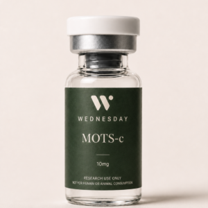 Metabolic Signaling Research
MOTS-c
10 mg View listing →
Metabolic Signaling Research
MOTS-c
10 mg View listing →
Frequently asked questions
What are mitochondrial-derived peptides?
They are short peptides encoded by small open reading frames within the mitochondrial genome, specifically inside the 12S and 16S ribosomal RNA genes. Known members are humanin, the six small humanin-like peptides (SHLP1–6) and MOTS-c. They are thought to act as signals from mitochondria to the rest of the cell.
What does humanin do?
In cell studies humanin protects neurons and other cells from apoptosis by binding the pro-apoptotic protein Bax and the growth-factor binding protein IGFBP-3, and by acting through cell-surface receptors. Its circulating levels decline with age in humans and mice. It has not been tested as a treatment in people.
What is the difference between humanin and MOTS-c?
Humanin is a 24-amino-acid peptide from the 16S rRNA gene that mainly regulates cell survival. MOTS-c is a 16-amino-acid peptide from the 12S rRNA gene that mainly regulates metabolism through AMPK and can move into the nucleus. Both decline with age in the models studied.
Are SHLPs the same as humanin?
No. The small humanin-like peptides are six distinct peptides from the same 16S rRNA region as humanin, described in 2016. SHLP2 and SHLP3 share some of humanin’s protective effects in cells, while SHLP6 promotes cell death. They are the least studied members of the family.
Have mitochondrial-derived peptides been tested in humans?
Only indirectly. Human studies have measured endogenous levels, showing decline with age and a rise in MOTS-c after exercise, and have examined genetic variants. The single interventional study used a modified MOTS-c analog in 20 participants for four weeks. No native MDP has been tested in a controlled human trial.
References & further reading
- Hashimoto Y, Niikura T, Tajima H, et al. A rescue factor abolishing neuronal cell death by a wide spectrum of familial Alzheimer’s disease genes and Aβ. Proc Natl Acad Sci U S A. 2001;98(11):6336–6341. doi:10.1073/pnas.101133498 / PMID 11371646
- Guo B, Zhai D, Cabezas E, et al. Humanin peptide suppresses apoptosis by interfering with Bax activation. Nature. 2003;423(6938):456–461. doi:10.1038/nature01627
- Lee C, Yen K, Cohen P. Humanin: a harbinger of mitochondrial-derived peptides? Trends Endocrinol Metab. 2013;24(5):222–228. doi:10.1016/j.tem.2013.01.005
- Lee C, Zeng J, Drew BG, et al. The mitochondrial-derived peptide MOTS-c promotes metabolic homeostasis and reduces obesity and insulin resistance. Cell Metab. 2015;21(3):443–454. doi:10.1016/j.cmet.2015.02.009 / PMID 25738453
- Cobb LJ, Lee C, Xiao J, et al. Naturally occurring mitochondrial-derived peptides are age-dependent regulators of apoptosis, insulin sensitivity, and inflammatory markers. Aging (Albany NY). 2016;8(4):796–809. doi:10.18632/aging.100943 / PMID 27070352
- Kim KH, Son JM, Benayoun BA, Lee C. The mitochondrial-encoded peptide MOTS-c translocates to the nucleus to regulate nuclear gene expression in response to metabolic stress. Cell Metab. 2018;28(3):516–524.e7. doi:10.1016/j.cmet.2018.06.008 / PMID 29983246
- Yen K, Mehta HH, Kim SJ, et al. The mitochondrial derived peptide humanin is a regulator of lifespan and healthspan. Aging (Albany NY). 2020;12(12):11185–11199. doi:10.18632/aging.103534 / PMID 32575074
- Reynolds JC, Lai RW, Woodhead JST, et al. MOTS-c is an exercise-induced mitochondrial-encoded regulator of age-dependent physical decline and muscle homeostasis. Nat Commun. 2021;12:470. doi:10.1038/s41467-020-20790-0
- Miller B, Kim SJ, Kumagai H, Yen K, Cohen P. Mitochondria-derived peptides in aging and healthspan. J Clin Invest. 2022;132(9):e158449. doi:10.1172/JCI158449 / PMID 35499074
- Ikonen M, Liu B, Hashimoto Y, et al. Interaction between the Alzheimer’s survival peptide humanin and insulin-like growth factor-binding protein 3 regulates cell survival and apoptosis. Proc Natl Acad Sci U S A. 2003;100(22):13042–13047. doi:10.1073/pnas.2135111100