Key takeaways
- A June 2026 study in Experimental Physiology reported that MOTS-c lowered fasting glucose and C-reactive protein and reduced NLRP3 inflammasome components in the hearts of type 2 diabetic rats.
- Three days later, a study in Inflammation and Regeneration from the Mayo Clinic and USC reported that MOTS-c activated AMPK in human mesenchymal stromal cells from obese donors but reduced their proliferation, increased senescence markers and blunted their repair effect in mice.
- The two findings are not contradictory so much as context-dependent: MOTS-c’s metabolic signaling appears robust, but its downstream consequences depend on the cell type and metabolic state it acts on.
- Together with three other June papers, this is an unusually productive month for MOTS-c research, and the negative result is arguably the most informative.
MOTS-c is a 16-amino-acid peptide encoded not in the nuclear genome but in the mitochondrial 12S ribosomal RNA gene, which makes it one of a small group of mitochondrial-derived peptides that act as signaling molecules between the organelle and the rest of the cell. Since its description in 2015, the literature has associated it with AMPK activation, improved glucose handling in mouse models, and a folate–methionine metabolic axis. June 2026 brought five new MOTS-c papers, including two primary studies whose conclusions appear, on first reading, to pull in opposite directions. One reports an anti-inflammatory and glucose-lowering effect in a rat model of type 2 diabetes. The other reports that the same peptide impairs the therapeutic function of human stem-like cells taken from people with obesity. Read carefully, they illustrate something important about how mitochondrial signaling peptides behave, and they are the most substantive additions to the MOTS-c record this year.
Study one: cardiac inflammasome in diabetic rats
Mills, de Souza, Pham and Mugisho, writing in Experimental Physiology on June 19, 2026, used a standard model of type 2 diabetes: rats fed a high-fat diet and given a low dose of streptozotocin to induce partial beta-cell loss. This produces hyperglycemia, insulin resistance and, over time, the systemic and cardiac inflammation that contributes to diabetic cardiomyopathy. Their target was the NLRP3 inflammasome, a multi-protein complex that processes interleukin-1β and interleukin-18 into their active forms and is a central driver of sterile inflammation in metabolic disease.1
In treated animals, fasting blood glucose and circulating C-reactive protein were significantly reduced. Plasma cytokines shifted selectively, interleukin-10, an anti-inflammatory signal, and interleukin-1β among them. In left ventricular tissue, immunohistochemistry showed lower levels of NLRP3 itself, of the adaptor protein ASC, and of cleaved caspase-1, the active enzyme that the assembled inflammasome produces. Correlation analyses linked interleukin-18 and interleukin-1β to low-density lipoprotein and uric acid, which the authors read as evidence that systemic metabolic health and cardiac inflammasome activity move together.1
The result extends earlier rodent work on MOTS-c and glucose metabolism into the cardiac inflammation that follows. Its limitations are those of the model: a chemically induced diabetes in rats, a single strain and sex, and outcomes measured by tissue staining and circulating markers rather than cardiac function. The authors frame it as suggesting a “novel therapeutic approach,” which is the appropriate register for a preclinical finding.
Study two: human stromal cells from obese donors
Xing and colleagues, in Inflammation and Regeneration on June 22, 2026, asked a different question. Mesenchymal stromal cells (MSCs) are being developed as cell therapies for kidney and cardiovascular disease, but MSCs from people with obesity are known to have impaired mitochondrial function and reduced repair capacity. Since MOTS-c is a mitochondrial signal that is depleted in obesity, the hypothesis was straightforward: restoring it might rescue the cells. The group, which includes Pinchas Cohen, whose laboratory first described MOTS-c, and Lilach Lerman at Mayo, isolated MSCs from abdominal adipose tissue of obese (BMI 30 or above) and lean donors, exposed them to MOTS-c in vitro, and then tested the pretreated cells in mice with renal artery stenosis.2
The metabolic arm of the hypothesis held: MOTS-c activated AMPK signaling in obese-donor MSCs, as expected. The functional arm did not. Treated cells showed reduced proliferation, increased expression of senescence genes, and elevated tumor necrosis factor-α. In vivo, MOTS-c pretreatment failed to improve kidney perfusion or fibrosis in the stenosis model, and it “blunted the reparative efficacy of lean MSCs”, that is, it made cells from lean donors, which normally work, work less well.2 The authors describe this as “metabolic–stemness uncoupling”: turning on the mitochondrial energy-sensing pathway did not restore, and in some respects impaired, the cells’ regenerative program.
The signal was the same in both studies; what differed was the cell that received it.
Reconciling the two
It is tempting to file one result as positive and the other as negative and move on. The more useful reading is that both are consistent with what is known about AMPK. In a diabetic animal, activating AMPK systemically pushes tissues toward glucose uptake and fatty-acid oxidation and away from the lipid-driven inflammation that feeds the NLRP3 inflammasome; the cardiac finding follows. In a cultured stromal cell whose job is to proliferate and secrete repair factors, sustained AMPK activation is a brake, it suppresses anabolic programs, including the mTOR-dependent growth that proliferation requires, and in already-stressed obese-donor cells that brake appears to tip them toward senescence.2
| Mills et al. Exp Physiol | Xing et al. Inflamm Regen | |
|---|---|---|
| Published | June 19, 2026 | June 22, 2026 |
| System | HFD/streptozotocin rat model of type 2 diabetes | Human adipose MSCs (obese and lean donors); mouse renal artery stenosis |
| Primary readouts | Fasting glucose, CRP, plasma cytokines, cardiac NLRP3/ASC/caspase-1 | Proliferation, senescence genes, TNF-α; kidney perfusion and fibrosis |
| AMPK-related signaling | Not the focus; metabolic improvement observed | Activated, as predicted |
| Functional outcome | Reduced systemic and cardiac inflammation markers | Reduced repair capacity; blunted lean-cell efficacy |
| Authors’ framing | Potential approach to diabetic cardiovascular risk | Mitochondrial signaling alone cannot reverse obesity-induced MSC dysfunction |
The two studies therefore describe two faces of one mechanism. MOTS-c reliably engages mitochondrial-to-nuclear metabolic signaling; whether that is helpful depends entirely on what the recipient cell is trying to do. For a whole animal under metabolic stress, the June data suggest a net anti-inflammatory shift. For a proliferating cell product, they suggest harm. Neither result generalizes to the other setting, and neither says anything about people.
The rest of June’s MOTS-c output
Three further papers rounded out the month. A group in Molecular Biology Reports reported that MOTS-c preserved bioenergetics in mitochondrial subpopulations and protected mitochondrial DNA integrity in a cardiac ischemia–reperfusion model.3 A study in Autophagy reported that MOTS-c reduced lysosomal membrane permeabilization and improved survival of transplanted soft tissue.4 And a review in the Journal of Translational Medicine made the case for MOTS-c as a candidate in inflammatory lung disease.5 The pattern across all five is consistent with the field’s trajectory: MOTS-c is being tested as a stress-response modulator in an expanding range of tissues, almost entirely in animal and cell models. Wednesday’s MOTS-c research overview and mitochondrial-derived peptides note place these in the longer arc.
Reading the evidence
No controlled human trial of MOTS-c has reported results. The human-cell study above used cultured cells, not people, and its in vivo component was in mice. MOTS-c was also one of seven peptides considered by FDA’s compounding advisory committee in July 2026, where the agency’s reviewers recommended against listing it; that discussion, covered in a separate note, was about pharmacy compounding and did not change the compound’s status as an unapproved substance.
Why the negative result matters most
Positive preclinical results for MOTS-c are not scarce. What the field has lacked is a well-designed study in which the peptide’s expected mechanism engaged and the expected benefit did not follow. The Xing paper supplies exactly that, from a group with every incentive to find the opposite. It identifies a boundary condition, metabolic state and cell type, that any future work with MOTS-c has to account for, and it argues against the assumption that “more mitochondrial signaling” is uniformly good. For researchers designing experiments with research-grade MOTS-c, that is more useful than another confirmation in a rodent model. It suggests that readouts should include proliferation and senescence markers alongside metabolic ones, and that donor or animal metabolic status should be treated as a variable rather than a nuisance.
MOTS-c is supplied by Wednesday for research use only. It is not an approved drug, no human trial has established any effect for it, and the June 2026 studies describe observations in rats, mice and cultured cells. Batch-specific certificates are in the COA library.
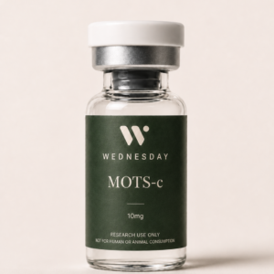 Metabolic Signaling Research
MOTS-c
Mitochondrial-derived peptide, research use only, third-party tested View listing →
Metabolic Signaling Research
MOTS-c
Mitochondrial-derived peptide, research use only, third-party tested View listing →
Frequently asked questions
What is MOTS-c?
MOTS-c is a 16-amino-acid peptide encoded within the mitochondrial 12S rRNA gene. It is one of several mitochondrial-derived peptides that signal from the organelle to the rest of the cell, and in animal and cell models it activates AMPK and influences glucose and lipid metabolism.
Does MOTS-c reduce inflammation?
In a June 2026 rat model of type 2 diabetes, MOTS-c reduced C-reactive protein and cardiac NLRP3 inflammasome components. In the same month, a study of human stromal cells from obese donors found it increased TNF-α and senescence markers. The effect on inflammation depends on the system, and there are no human data.
What did the 2026 Mayo Clinic MOTS-c study find?
That MOTS-c activated AMPK in mesenchymal stromal cells from obese donors but reduced their proliferation, raised senescence gene expression and elevated TNF-α, and that pretreating cells with MOTS-c blunted their ability to repair kidney injury in mice. The authors called this metabolic–stemness uncoupling.
Has MOTS-c been tested in humans?
No controlled human trial of MOTS-c has published results. All evidence to date comes from rodent models and cultured cells, including human cells studied in vitro. MOTS-c is not an approved drug.
Is MOTS-c approved for compounding?
No. An FDA advisory committee voted 7–5 in July 2026 to recommend it for consideration on the 503A compounding list, against the recommendation of FDA’s own reviewers. The vote is non-binding and no rule has been issued.
References & further reading
- Mills AR, de Souza A, Pham T, Mugisho OO. Mitochondrial peptide MOTS-c suppresses systemic and cardiac inflammasome activation in a diabetic rat model. Exp Physiol. 2026. doi:10.1113/EP093714
- Xing L, Lu B, Zhu X, Al Saeedi M, Lerman A, Eirin A, Cohen P, Lerman LO. Mitochondrial-derived peptide MOTS-c activates metabolic signaling but blunts reparative function in human mesenchymal stromal cells. Inflamm Regen. 2026. doi:10.1186/s41232-026-00431-7
- Santhanam SS, Jayaraman S, Rajesh SS, et al. MOTS-c preserves mitochondrial subpopulation bioenergetics and genome integrity to attenuate cardiac ischemia reperfusion injury. Mol Biol Rep. 2026. doi:10.1007/s11033-026-12064-7
- Shi J, Wu Y, Liu X, et al. MOTS-c, a mitochondrial-derived peptide, ameliorates lysosomal membrane permeability and improves survival of soft tissue transplantation. Autophagy. 2026. doi:10.1080/15548627.2026.2677180
- Amado CA, Agüero J, García-Unzueta M, et al. MOTS-c: How a secreted mitochondrial microprotein may become a potential treatment for inflammatory lung diseases. J Transl Med. 2026. doi:10.1186/s12967-026-08398-2
- Lee C, Zeng J, Drew BG, et al. The mitochondrial-derived peptide MOTS-c promotes metabolic homeostasis and reduces obesity and insulin resistance. Cell Metab. 2015;21(3):443–454. doi:10.1016/j.cmet.2015.02.009
- U.S. Food and Drug Administration. FDA Briefing Document, Pharmacy Compounding Advisory Committee Meeting, July 23–24, 2026. fda.gov (PDF)